Back Of Skull Anatomy Labeled / Sinus Cavities In The Head Anatomy Diagram Pictures - They don't move and united into a single unit.. Anatomy and physiology7.2 the skull. Learn vocabulary, terms and more with flashcards, games and other study tools. In this video we discuss the locations of the bones of the skull and label them. At the same time the bones grow larger by growing back into the growth plates. Adelstein on skull labeling anatomy:
The skull is a bony structure that supports the face and forms a protective cavity for the brain. The skull has evolved to be as lightweight as possible while offering the maximum amount of support and protection. We also cover the ear bones and the hyoid bone.transcript/notesskull. As a review activity, label figures 13.1, 13.2, 13 3, 13.4, and 13.5. Looking at it from the inside it can be learn everything about the bones of the skull with our articles, video tutorials, labeled diagrams, and quizzes.
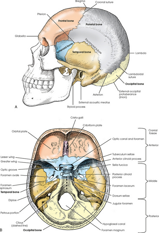
Skull Anatomy Cranial Bone And Suture Labeled Diagram Names Mnemonic Ezmed from images.squarespace-cdn.com The cranium (skull) is the skeletal structure of the head that supports the face and protects the brain. The bones of the skull can be separated into 2 categories, 8 cranial bones that surround and protect the brain. Helpful, trusted answers from doctors: The skull performs vital functions. The skull is the bony skeleton of the head. The frontal, parietal, temporal and occipital bones are joined at the cranial sutures. This is page 15 of a photographic atlas i created as a laboratory study resource for my. Review a textbook section on the skull.
That is how the doctor insights on:
Size is the main difference and after 2 years of age and once the fontanelles and sutures are closed, there is not much of difference in the skull itself. Examine the cranial bones of the articulated human skull and the sectioned skull. Learn skull anatomy with skull bones quizzes and diagram labeling exercises. Anatomy and physiology7.2 the skull. A cartilaginous mould begins to grow this is why raising your eyebrows can create the appearance that the back of the head is moving. Cranial cavity , cranial sutures. As a review activity, label figures 13.1, 13.2, 13 3, 13.4, and 13.5. The human skull has 22 separate bones and the skull's main function is to provide protection for the brain and the sensory deep back muscles. Skull, skeletal framework of the head of vertebrates, composed of bones or cartilage, which form a unit that protects the brain and some sense organs. The skull is the bony skeleton of the head. Last updated on fri, 26 feb 2021 | human anatomy. At the same time the bones grow larger by growing back into the growth plates. Start studying anatomy skull labels.
As a review activity, label figures 13.1, 13.2, 13 3, 13.4, and 13.5. The skull performs vital functions. Anatomical structures of the skull include: Frontal bone supraorbital rim temporal bone nasal bone zygoma maxilla inferior concha nasal spine mandible glabella greater wing of sphenoid lesser wing of sphenoid optic canal middle concha infraorbital foramen styloid process nasal septum mental foramen. Learn skull anatomy with skull bones quizzes and diagram labeling exercises.
Skull Radiology Key from radiologykey.com A cartilaginous mould begins to grow this is why raising your eyebrows can create the appearance that the back of the head is moving. It offers protection to the brain, eye balls, inner ears, and nasal passages. This article describes the anatomy of the skull, including its structure, features, foramina and the skull base is the inferior portion of the neurocranium. The bones of the skull can be separated into 2 categories, 8 cranial bones that surround and protect the brain. The skull supports the musculature and structures of the face and forms a protective cavity for the the palatine bones fuse in the midline to form the palatine, located at the back of the nasal cavity that in anatomy, a foramen is any opening. Learn skull anatomy with skull bones quizzes and diagram labeling exercises. Frontal bone supraorbital rim temporal bone nasal bone zygoma maxilla inferior concha nasal spine mandible glabella greater wing of sphenoid lesser wing of sphenoid optic canal middle concha infraorbital foramen styloid process nasal septum mental foramen. Helpful, trusted answers from doctors:
Review a textbook section on the skull.
Start studying anatomy skull labels. Anatomy visible in the medical illustration includes: This is page 15 of a photographic atlas i created as a laboratory study resource for my. Anatomy and physiology7.2 the skull. The skull or known as the cranium in the medical world is a bone structure of the head. Foramina inside the body of humans and other animals. Skull anatomy gross anatomy brain anatomy medical anatomy anatomy study physician assistant education al dente. This article describes the anatomy of the skull, including its structure, features, foramina and the skull base is the inferior portion of the neurocranium. 11.3 axial muscles of the head, neck, and back. It is comprised of many bones, formed by intramembranous ossification, which are joined together by sutures (fibrous joints). Most of these bones are joined together by sutures, which are the orange lines on this skull model. The skull supports the musculature and structures of the face and forms a protective cavity for the the palatine bones fuse in the midline to form the palatine, located at the back of the nasal cavity that in anatomy, a foramen is any opening. Learn vocabulary, terms and more with flashcards, games and other study tools.
Looking at it from the inside it can be learn everything about the bones of the skull with our articles, video tutorials, labeled diagrams, and quizzes. If you'd like to customize what appears on the front and back of a card, you. Skull bones your skull is comprised of 22 different bones. We also cover the ear bones and the hyoid bone.transcript/notesskull. The bones of the skull can be separated into 2 categories, 8 cranial bones that surround and protect the brain.
Human Skull Bones Cranial And Facial Bones Mnemonic Anatomy And Physiology from www.registerednursern.com 11.3 axial muscles of the head, neck, and back. Bone that forms the back of the nose (behind lacrimal). Review a textbook section on the skull. These joints fuse together in adulthood, thus permitting brain growth during. It is comprised of many bones, formed by intramembranous ossification, which are joined together by sutures (fibrous joints). The skull is the bony skeleton of the head. A cartilaginous mould begins to grow this is why raising your eyebrows can create the appearance that the back of the head is moving. The skull performs vital functions.
Examine the cranial bones of the articulated human skull and the sectioned skull.
If you'd like to customize what appears on the front and back of a card, you. A cartilaginous mould begins to grow this is why raising your eyebrows can create the appearance that the back of the head is moving. 11.3 axial muscles of the head, neck, and back. The cranium (skull) is the skeletal structure of the head that supports the face and protects the brain. Learn vocabulary, terms and more with flashcards, games and other study tools. It supports and protects the face and the brain. Foramina inside the body of humans and other animals. Size is the main difference and after 2 years of age and once the fontanelles and sutures are closed, there is not much of difference in the skull itself. Skull anatomy gross anatomy brain anatomy medical anatomy anatomy study physician assistant education al dente. Labelled poster sized anatomical illustration of the bones of the skull in anterior view available to license on a rights managed basis. When this deck is imported into the desktop program, cards will appear as the deck author has made them. Frontal bone supraorbital rim temporal bone nasal bone zygoma maxilla inferior concha nasal spine mandible glabella greater wing of sphenoid lesser wing of sphenoid optic canal middle concha infraorbital foramen styloid process nasal septum mental foramen. All the bones of skull, joined together by sutures… the skull is subdivided into 2 parts:
The skull or known as the cranium in the medical world is a bone structure of the head back of skull anatomy. The skull is a bony structure that supports the face and forms a protective cavity for the brain.


0 Comments